





Medical Articles
Explore expert medical articles on veterinary radiology, AI-powered diagnostics, and the latest advancements in clinical imaging.
Filter by categories

Featured
Getting the Most Out of Skull Radiographs in Dogs and Cats
Skull radiographs can be a valuable tool when images are obtained with precison. While the anatomy is complex and positioning…


Dog CCL Tear on Radiographs: Cranial Cruciate Ligament Rupture, Joint Effusion, and Surgical Planning
Learn how radiographs support diagnosis and surgical planning for dog CCL tear cases, including CCL rupture, stifle effusion, osteophytes, and…

Periodontal Disease in Dogs: Signs, Causes, and What Practitioners Frequently Miss
Learn how dental radiographs reveal periodontal disease in dogs, including alveolar bone loss, furcation exposure, staging, and treatment planning.


Pyometra in Dogs: Radiographic Diagnosis, Distinguishing Open vs. Closed, and When to Act Fast
Pyometra is one of the more common life-threatening emergencies that presents to the general practitioner. Prompt diagnosis is essential, as…

Tracheal Collapse in Dogs: Signs, Causes, and the Limits of Static Imaging
Tracheal collapse in dogs is a common cause of chronic cough, especially in toy and small-breed patients. It is often…

5 Tips for Evaluating the Feline Heart on Radiographs
Evaluating the feline on radiographs takes a different approach than evaluating the canine heart. Cats can have clinically meaningful heart…

Hip Dysplasia in Dogs: Radiographic Diagnosis, Clinical Signs, and Treatment Planning
Hip dysplasia in dogs is one of the most important orthopedic diseases seen in general practice, especially in large and…

Abdominal Mass Effect vs. Mid Abdominal Mass: What’s the Difference?
In veterinary radiology, small wording differences can make a meaningful clinical difference. One example is the distinction between abdominal mass…


Cat Constipation: Radiographs, Megacolon, and When Constipation Becomes an Emergency
Cat constipation is common, but it should not be treated as a single, simple problem. Some cats have occasional hard…

Canine Heart Disease Stages and the Role of Radiographs in Long-Term Management
Canine Heart Disease Stages and the Role of Radiographs in Long-Term Management Canine heart disease stages are not just diagnostic…

Cat Linear Foreign Body: Radiographic Findings, Diagnosis, and When Surgery Is Needed
Linear foreign bodies in cats, such as string or yarn, can anchor under the tongue or in the stomach and…

Signs of intestinal obstruction in dogs: Radiographs, Diagnosis and When Surgery Is Needed
Intestinal obstruction in dogs is a common emergency that can quickly become life-threatening. Learn the signs of intestinal obstruction in…


Dog Bone Cancer Symptoms: Radiographic Clues and Early Detection of Osteosarcoma in Dogs
Dog bone cancer symptoms in dogs often start with progressive lameness, pain, swelling, and poor response to NSAIDs. Learn how…
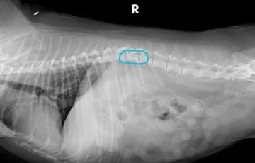
IVDD in Dogs: How Radiographs, CT, and MRI Guide Diagnosis and Treatment Decisions
A clinical guide to IVDD in dogs: neurologic grading, what spinal radiographs show and miss, when to escalate to CT…

Radiographic Signs of Canine Cardiac Disease: VHS, VLAS, and When to Use Echocardiography
Learn how to identify cardiac enlargement on canine radiographs using VHS and VLAS, recognize pulmonary changes, and determine when echocardiography…

Canine Thoracic Radiograph Interpretation: A Systematic Approach for General Practitioners
How to identify, characterize, and stage pulmonary masses and nodules on canine radiographs, and when radiographs are enough versus when…

Canine Lung Cancer on Radiographs: Primary Tumors, Metastatic Disease, and When to Push for CT
How to identify, characterize, and stage pulmonary masses and nodules on canine radiographs, and when radiographs are enough versus when…

Megaesophagus in Dogs: Radiographic Diagnosis, Aspiration Pneumonia Risk, and Clinical Management
Learn how to diagnose megaesophagus in dogs on radiographs, recognize aspiration pneumonia risk, investigate underlying causes, and manage canine megaesophagus…

Radiology Case of the Week | Canine Pleural Effusion
Patient History The patient is a 5-year-old MC mixed breed dog with a diagnosis of idiopathic chylothorax since February 2020…


Pleural Effusion in Cats: Radiographic Diagnosis, Differential Diagnosis by Signalment, and Triage Decisions
Pleural effusion in cats is a major cause of respiratory distress. Learn the key radiographic signs, signalment-based differentials, and how…

Diaphragmatic Hernia in Dogs and Cats: Diagnosis, Imaging Strategies, and the Impact of AI
A diaphragmatic hernia occurs when abdominal viscera protrude through a defect in the diaphragm into the thoracic cavity. In small…

Uterine Distension in Dogs and Cats: Detection, Interpretation, and the Clinical Value of AI
Uterine distension in dogs and cats can indicate serious reproductive disease such as pyometra, mucometra, or metritis. Learn how to…

Thoracic Masses in Dogs and Cats: Diagnostic Approach, Imaging Strategies, and the Role of AI
This article explores the major causes of thoracic masses, how to localize them radiographically, when to pursue advanced diagnostics, and…

Increased Thoracic Opacities
Increased thoracic opacity on radiographs can indicate pulmonary disease, thoracic masses, or pleural effusion in dogs and cats. This guide…

Bone Lesions vs. Artifacts
Distinguishing true bone lesions from artifacts is one of the most common and critical challenges in veterinary radiology.

The Holiday GDV Surge
We found a clear and concerning trend: GDV cases increase significantly during the holidays, including Thanksgiving, Christmas, and the Fourth…

Esophageal Foreign Bodies in Dogs and Cats: Early Detection, Imaging Strategies, and the Role of AI
Esophageal foreign bodies (EFBs) are a common and potentially life-threatening emergency in veterinary medicine.

Be Specific, Get Better Answers
When requesting a STAT consultation, every detail you provide matters. A clear clinical history and well-defined primary concern help the…

Linear Foreign Bodies in Dogs and Cats: Diagnostic Strategies, Radiographic Clues, and the Role of AI
Linear foreign bodies (LFBs) represent a particularly dangerous subset of gastrointestinal emergencies in veterinary medicine. Often caused by the ingestion…
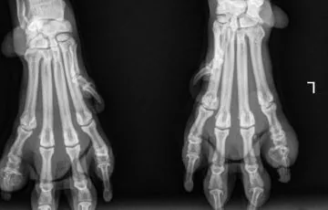
Lysis of the Third Phalanx: Why Early Detection Matters and How Al Can Help
Lysis of the third phalanx also referred to as P3 bone loss characterised by destruction or resorption of this bone…

Left Atrial Enlargement in Dogs and Cats: Detection, Interpretation, and the Clinical Value of AI
Left atrial enlargement (LAE) is a common but important radiographic and echocardiographic finding in small animal cardiology. LAE commonly reflects…

Abdominal Masses in Dogs and Cats: Detection, Diagnostic Strategies, and the Role of AI
The discovery of an abdominal mass in dogs and cats is a clinically significant finding. These masses can arise from…

Bladder Stones in Dogs: What Multi-Year Data from SignalPET Reveals About Risk by Region, Breed, and Age
Bladder stones are among the most frequently encountered urinary conditions in veterinary practice. While most clinicians are familiar with their…

Urethral Calculi in Dogs and Cats: Radiographic Detection, Interpretation Tips, and the Role of AI
Urethral calculi, also known as urethral stones, are a common health concern in both dogs and cats.

A Review of Diaphragmatic Hernias
Diaphragmatic hernias are commonly recognized by small animal clinicians and may be congenital or traumatic in origin, and acute or…

Guidelines for optimal use of dental radiography
Dental radiography is fast becoming the standard of care as part of a COHAT (comprehensive oral health assessment and treatment).…

Festive Follies – Tis the season for esophageal foreign bodies!
As we get to crack open the wine and tuck into the delicacies, we wanted to reflect on some common…

Radiology Case of the Week | Canine Neck Swelling
A geriatric, male intact pug was taken in by a rescue organization. He presented to the veterinarian for evaluation of…

Radiology Case of the Week | Canine Appendicular Bone Fracture
Radiology Case of the Week | Canine Pleural Effusion Patient History Presumed trauma (hit by car) and left pelvic limb…

Radiology Case of the Week | Canine Gastrointestinal Foreign Body
Canine gastrointestinal foreign bodies occur when ingested objects become lodged within the stomach or intestines, potentially causing obstruction and gastrointestinal…
Radiology Case of the Week | Feline Miliary Pulmonary Pattern
In this Week's Radiology Case, we take a dive into Feline Miliary Pulmonary Pattern, going over the radiographic appearance and…
Radiology Case of the Week | Feline Bronchiectasis
Feline bronchiectasis is a chronic airway condition characterized by permanent dilation of the bronchi due to ongoing inflammation or infection.…

Radiology Case of the Week | Canine Esophageal Foreign Body
Patient History Unknown Radiographs Radiology Test Results What’s Abnormal about this Case? In this week’s case, the lesion is not…

Radiology Case of the Week | Feline Congenital Thoracic Lordosis
Radiology Case of the Week | Canine Pleural Effusion Patient History An 8 week old MI DSH presented to the…
Radiology Case of the Week | Canine Pleural Effusion
Patient History The patient is a 5-year-old MC mixed breed dog with a diagnosis of idiopathic chylothorax since February 2020…

Radiology Case of the Week | Peritoneal Pericardial Diaphragmatic Hernias
Peritoneal-pericardial diaphragmatic hernia (PPDH) is a congenital condition whereby the normal anatomic separation between the pericardium and peritoneal cavity fails…

Radiology Case of the Week | Feline Radial Hemimelia
Recently, we have seen two cases of feline radial hemimelia, in which the radius is incompletely formed or absent.

