



6 Types of Veterinary Imaging Equipment and When to Use Each
Veterinary imaging equipment ranges from first-line digital radiography to advanced CT and MRI. Learn what each modality shows best, when it may be recommended, and why different imaging methods are often used together.

6 Types of Veterinary Imaging Equipment and When to Use Each

Veterinary diagnostic imaging allows clinicians to evaluate anatomy and disease that cannot be fully assessed through physical examination or laboratory testing alone. However, each type of veterinary imaging equipment provides different information.
Digital radiography is often the first step for evaluating bones, the thorax, and the abdomen. Veterinary ultrasound provides real-time soft-tissue imaging, while CT and MRI offer detailed cross-sectional views for more complex cases. Dental radiography and fluoroscopy address specialized clinical questions that standard radiographs may not answer.
The appropriate modality depends on the suspected condition, body region, required detail, urgency, availability, cost, and the patient’s ability to tolerate positioning, sedation, or anesthesia.
Key Takeaways
- Each imaging modality has specific strengths and limitations.
- Digital X-ray is commonly used as a fast, accessible first-line test.
- Ultrasound is valuable for evaluating soft tissues, organs, fluid, and blood flow.
- CT and MRI provide cross-sectional imaging but answer different clinical questions.
- Dental X-rays and fluoroscopy evaluate structures or functions that may not be visible on standard radiographs.
- Different imaging modalities are often complementary rather than interchangeable.
Veterinary Imaging Equipment Comparison

1. Digital X-Ray: A First-Line Tool for Bones, the Chest, and the Abdomen

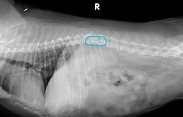
Digital veterinary X-ray equipment uses ionizing radiation to create two-dimensional images. Radiographs are commonly selected as an initial imaging test because they are fast, accessible, and useful across several body regions.
Common indications include fractures, joint disease, bone lysis or proliferation, pulmonary abnormalities, changes in cardiac silhouette size, pleural fluid or gas, gastrointestinal obstruction, abdominal organ enlargement, mineralized calculi, and radiopaque foreign material.
Thoracic radiographs help assess the lungs, pleural space, mediastinum, diaphragm, and cardiac silhouette. They may be used during the investigation of conditions such as canine lung cancer and canine cardiac disease.
Abdominal radiographs provide an overview of organ size, position, gas distribution, and serosal detail. They may reveal a mass effect, although ultrasound or CT is often needed to identify its organ of origin. Learn more in SignalPET’s guide to abdominal masses in dogs and cats.
The principal limitation of radiography is superimposition. Multiple structures are projected onto the same image, which may obscure subtle abnormalities. Correct positioning and orthogonal views are therefore important.
2. Ultrasound: Real-Time Imaging of Soft Tissues and Internal Organs

Veterinary ultrasound uses high-frequency sound waves to create real-time images without ionizing radiation. It is particularly useful for examining internal organ architecture, fluid, movement, and blood flow.
Ultrasound may be used to assess the liver, spleen, kidneys, urinary bladder, gastrointestinal tract, pancreas, adrenal glands, reproductive organs, abdominal masses, and free abdominal or thoracic fluid.
Echocardiography is a specialized form of ultrasound used to evaluate cardiac chambers, valves, contractility, and blood flow. Thoracic radiographs and echocardiography provide complementary information: radiographs show overall cardiac size and associated pulmonary changes, while echocardiography evaluates internal cardiac structure and function.
Ultrasound can also guide fine-needle aspiration, biopsy, or fluid drainage. Its limitations include operator dependence and reduced effectiveness through bone or gas-filled structures. A normal or inconclusive ultrasound examination may not completely exclude disease.
3. Computed Tomography: Detailed Cross-Sectional Imaging for Complex Cases

A veterinary CT scan uses X-rays acquired from multiple angles to create cross-sectional images. These images can be reviewed in different planes or reconstructed into three-dimensional models.
CT is especially useful for complex fractures, skull and facial trauma, nasal disease, pulmonary nodules, thoracic masses, polytrauma, tumor assessment, and surgical planning.
Unlike conventional radiographs, CT reduces superimposition and provides more precise anatomic localization. It may identify small pulmonary lesions or show how a mass relates to nearby vessels, airways, bones, or organs.
Intravenous contrast may be administered to evaluate vascular structures, tissue enhancement, or tumor margins. Sedation or general anesthesia is often required because patient movement reduces image quality.

Schedule a Demo
Ready to discover how SignalPET is designed to help you leverage radiology in your practice?
4. Magnetic Resonance Imaging: Advanced Evaluation of the Brain, Spine, and Soft Tissues

Veterinary MRI uses a strong magnetic field and radiofrequency energy rather than ionizing radiation. It provides excellent contrast between different soft tissues.
MRI is frequently used to evaluate brain disease, seizures with suspected structural causes, spinal cord compression, intervertebral disc disease, nerve roots, peripheral nerves, and selected musculoskeletal or soft-tissue abnormalities.
Different MRI sequences can emphasize fluid, fat, hemorrhage, inflammation, and contrast enhancement. This allows the examination to be adapted to the suspected condition.
MRI studies usually take longer than CT examinations and are highly sensitive to movement, so general anesthesia is commonly required. MRI is not universally superior to CT. CT is often preferred for lungs, bone, mineralization, and certain trauma cases, while MRI is generally more informative for the brain, spinal cord, and nerves.
5. Dental X-Ray: Detecting Disease Below the Gumline

A visual oral examination cannot assess the full tooth root or supporting periodontal bone. Veterinary dental X-rays are intraoral radiographs obtained using a small sensor or plate positioned inside the mouth.
They can identify periodontal bone loss, root fractures, periapical disease, unerupted teeth, retained root fragments, feline tooth resorption, and jaw fractures.
Dental radiographs are particularly important before and after extractions. They help assess the number and shape of the roots, the surrounding bone, and possible complications, and can confirm whether the entire root has been removed.
Because accurate intraoral positioning requires the patient to remain still, dental radiographs are usually obtained under general anesthesia.
6. Fluoroscopy: Real-Time Imaging of Motion and Function

Veterinary fluoroscopy uses continuous or pulsed X-rays to create moving images. It is useful when the clinical question involves movement or function rather than static anatomy.
Common applications include swallowing studies, assessment of tracheal collapse, esophageal motility, gastrointestinal motility, dynamic orthopedic evaluation, and image-guided catheter or stent placement.
A fluoroscopic swallowing study may show where a bolus is delayed, misdirected, or aspirated. These abnormalities may not be captured on a standard radiograph.
Radiation safety is especially important because veterinary personnel may remain close to the patient during image acquisition. Exposure time, collimation, distance, protective equipment, and procedural planning should be carefully managed.
How to Choose the Right Veterinary Imaging Modality
Modality selection should be guided by the clinical question.
Radiographs may be sufficient for a straightforward fracture, while CT may be needed for a complex articular injury. Ultrasound is often appropriate for abdominal organs and fluid, while MRI is generally preferred for the brain or spinal cord.
Additional considerations include patient stability, the required level of detail, the need for sedation or anesthesia, availability, urgency, cost, and whether image-guided sampling is required.
Imaging studies are often sequential. Radiographs may identify an abnormality, ultrasound may characterize it, and CT or MRI may define its full extent before surgery or treatment.
Practices also need a reliable way to manage studies after they have been acquired. SignalPET PACS supports the storage, viewing, organization, comparison, and sharing of imaging studies from multiple modalities, including radiographs, ultrasound, and CT. When specialist interpretation is needed, SignalPET STAT and Radiologist Report services can support eligible urgent or routine cases involving modalities beyond radiographs, including ultrasound and CT.
How SignalPET Supports Veterinary Imaging Workflows
AI-assisted radiograph analysis can provide an additional review layer for eligible X-ray studies. It may help draw attention to clinically meaningful abnormalities and support a systematic assessment of the available radiographic views.
SignalPET’s AI interpretation capabilities support veterinary radiography specifically. They should not be interpreted as automated analysis of every modality discussed in this article. Ultrasound, CT, MRI, and fluoroscopy require modality-specific acquisition and interpretation.
Other parts of the SignalPET platform support broader imaging workflows. SignalPET PACS can be used to store, access, organize, compare, and share imaging studies from multiple modalities, including radiographs, ultrasound, and CT. SignalPET STAT and Radiologist Report services can also support eligible ultrasound and CT cases when specialist interpretation is required.
These services perform different roles:
- SignalPET AI provides an additional review layer for eligible radiographic studies.
- SignalPET PACS supports the management and viewing of multimodality imaging studies.
- SignalPET STAT provides expedited specialist interpretation for eligible urgent cases.
- SignalPET Radiologist Reports provide specialist-reviewed interpretation when a formal imaging report is required.
AI and specialist services support rather than replace veterinarian judgment. Imaging findings must still be correlated with the patient’s history, physical examination, laboratory results, image quality, and complete clinical picture.

FAQs
Digital radiography is among the most commonly used modalities because it is fast, accessible, and useful for evaluating bones, the thorax, and the abdomen. The appropriate first test still depends on the patient’s clinical signs and suspected condition.
X-rays create two-dimensional projection images and are particularly useful for bones, lungs, and broad anatomic patterns. Ultrasound uses sound waves to evaluate soft-tissue organs, fluid, movement, and blood flow in real time.
CT may be recommended when radiographs do not provide sufficient anatomic detail. Common indications include complex fractures, skull or nasal disease, pulmonary nodules, major trauma, tumor assessment, and surgical planning.
Many radiographs and ultrasound examinations can be performed without anesthesia. CT, MRI, and dental radiography more commonly require sedation or general anesthesia to prevent motion and obtain diagnostic-quality images.
The answer depends on the tissue. Ultrasound is often used for abdominal organs, fluid, and cardiac structures. MRI generally provides greater detail for the brain, spinal cord, nerves, and many other soft tissues.
Table of Contents


Book a Demo
A New Standard of Care: All of your patients now benefit from an additional opinion, without ever leaving your office
SignalPET helps you bring real diagnostic expertise to All of your cases with three layers of support.